
Citicoline: A potential candidate drug for controlling myopia
The current methods for controlling the progression of myopia mainly include optical correction, drug intervention and refractive surgery.
In terms of drug treatment, atropine is a commonly studied option.
It can reduce the progression of myopia by approximately 90%, but it also has side effects such as blurred vision when looking at close objects and photophobia.
Researchers have been exploring new drug targets.
This study, published in "Biomedicines", indicates that choline phosphocholine can increase the level of dopamine in the retina, thereby inhibiting the progression of myopia.
mechanism of action of citicoline
Citicoline is a substance that participates in the dopamine pathway and has shown beneficial effects in the treatment of glaucoma and amblyopia.
Dopamine is the main neurotransmitter in the retina of mammals and plays a crucial role in regulating the growth of the eyeball.
When the transmission of visual signals decreases, the dopamine levels in the retina will drop, which may trigger the signal for excessive growth of the eyeball.
Citicoline can activate the biosynthesis pathway of phospholipids in the neuronal membrane structure and stimulate multiple neurotransmitter systems, including the dopamine system.
Research Methodology
This study employed a rat model of induced myopia through lens implantation.
The myopia was induced by suturing an S-10.00D concave lens around the right eye socket of the rats.
The experiment was divided into several groups:
Negative control group: No wearing of glasses, no administration of medication
Positive control group: Wearing glasses, no medication given
Treatment group: Wearing glasses, they were given 50, 100, 200 or 300 mg/kg/day of cytidine diphosphate choline respectively.
The administration method was oral gavage, and it lasted for 7 days.
After the experiment, the researchers removed the eyeballs of the rats, measured the eye axis length, and evaluated the changes in the scleral tissue using immunohistochemistry and tissue staining methods.
Change in length of eyeball
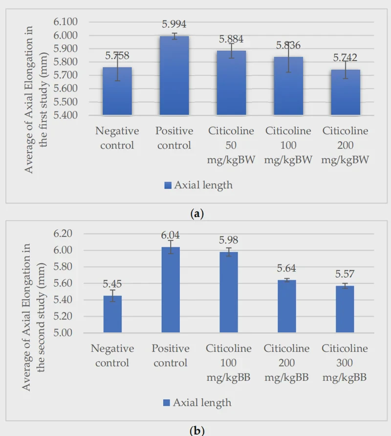
The positive control group showed a significant increase in the eye axis after wearing the lenses, indicating that the myopia model was successfully induced.
After administration of citicoline, the axial lengths of the eyes in the 100mg/kg group and the 200mg/kg group were significantly shorter than those in the positive control group.
Especially in the 200mg/kg group, the eye axis length was the closest to that of the negative control group.
This indicates that choline phosphocholine can effectively inhibit the abnormal elongation of the eye axis caused by lens-induced factors.
expression changes of MMP-2
MMP-2 is an enzyme that can degrade the extracellular matrix of the sclera.
Its excessive activity can lead to the thinning and weakening of the sclera.
In the immunohistochemical staining, the scleral tissue of the positive control group (B) showed a large amount of dark brown MMP-2 positive staining, indicating that the expression level of this enzyme was very high.
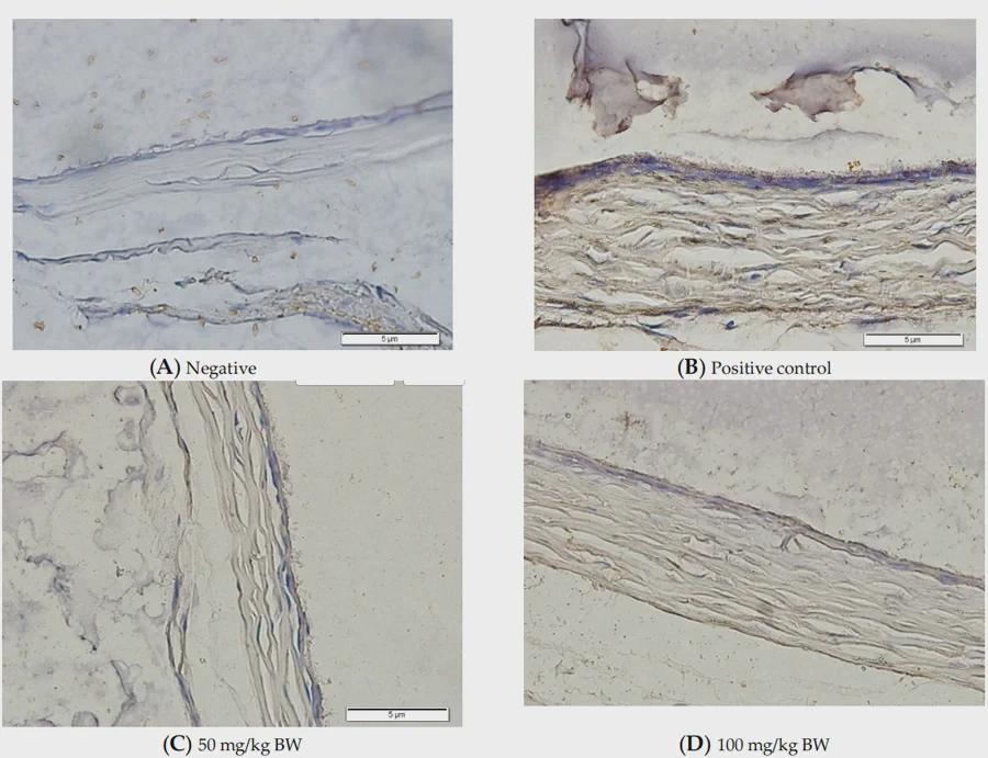
The brown staining in the negative control group (A) and the treatment group was significantly reduced.
The statistical results showed that the mean expression of MMP-2 in the positive control group was 70.88%, while it was only 18.38% in the 200mg/kg treatment group. The difference was statistically significant.
This indicates that choline phosphocholine can effectively inhibit the excessive activity of MMP-2 in the sclera, thereby reducing the damage to the scleral structure.
Changes in thickness of sclera
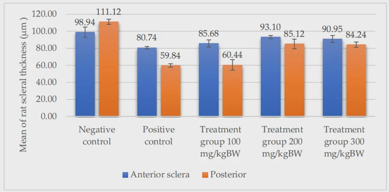
The study also measured the thickness of the anterior and posterior poles of the sclera.
The results showed that the sclera in the positive control group was significantly thinner, especially in the posterior pole area, which was consistent with the characteristics of scleral remodeling during the development of myopia.
After administration of 200mg/kg and 300mg/kg of choline phosphocholine, the scleral thickness in both the anterior and posterior regions was significantly higher than that of the positive control group.
Specifically, in the anterior region, the 200mg/kg group reached 93.10μm (compared to 80.74μm in the positive control group), and in the posterior region, it reached 85.12μm (compared to 59.84μm in the positive control group).
This indicates that choline phosphocholine is helpful in maintaining or restoring the thickness of the sclera.
expression changes of TGF-β1
TGF-β1 is a cytokine that promotes the synthesis of extracellular matrix in scleral cells and plays a crucial role in maintaining the structure and strength of the sclera.
The research found that the intensity of TGF-β1 positive staining in the 300mg/kg citicoline group was significantly higher than that in the other groups.
The statistical results show that the expression level of TGF-β1 in the anterior pole of the 300mg/kg group reached 12.86%, which was much higher than the 3.34% of the positive control group.
This indicates that higher doses of choline phosphocholine can promote the expression of TGF-β1, which is beneficial for the repair process of the sclera.
changes in fibroblast proliferation
Ki-67 is a marker that reflects the proliferative activity of cells.
Its expression level can indicate the degree of division activity of scleral fibroblasts.
At the posterior pole of the sclera, the Ki-67 expression in the positive control group was 6.23%, significantly higher than that in the negative control group (3.53%), indicating that the cell proliferation activity in the posterior pole of the sclera was enhanced after myopia induction.
After administration of 200mg/kg choline bitartrate, the expression of Ki-67 decreased to 4.64%, approaching the normal level.
This indicates that choline phosphocholine helps restore the balance of scleral cell proliferation.
Summary:
Citicoline can reduce the expression level of MMP-2 in the sclera, increase the thickness of the sclera, and a higher dose (300mg/kg) of citicoline can significantly enhance the expression of TGF-β1, which is helpful in initiating the repair process of the sclera and regulating the proliferation activity of scleral fibroblasts.
This study provides new experimental evidence for the potential use of citicoline as a drug for the intervention of myopia.
The current conclusion is still based on animal models, and the specific details of its mechanism of action and its effects in humans still require further research for verification.
Source: https://doi.org/10.3390/biomedicines10102600
Related products


Related news
-
Citicoline improves your brain function - cerebrovascular disease
2024-09-29 -
Citicoline - brain metabolic activator
2024-12-23 -
therapeutic effect of citicoline in treating severe and remission stages of Parkinson disease
2026-05-12 -
Will there be any side effects take too much Citicoline Sodium?
2026-06-15
