
Glutathione is crucial for immune response mediated by NK cells
Natural killer (NK) cells are of vital importance for immune protection against tumors and viruses.
The disease environment can cause oxidative stress, which damages the functions of immune cells.
Glutathione (GSH) is a major cellular antioxidant that is crucial for the immune response. However, the way it regulates the function of NK cells remains largely unknown.
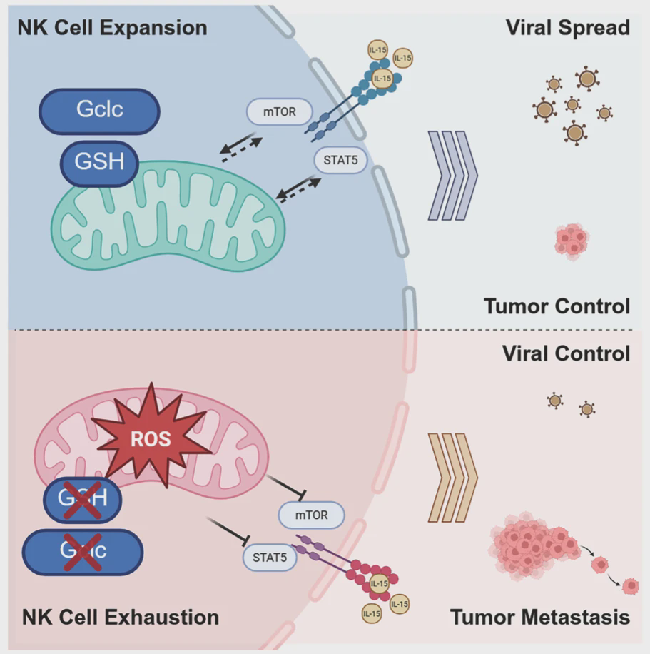
Using a mouse model with specific deletion of the catalytic subunit of glutamate-cysteine ligase (Gclc) in NK cells, it was demonstrated that glutathione supports the activation of NK cells driven by interleukin-15 (IL-15).
The deficiency of Gclc will lead to the accumulation of reactive oxygen species (ROS) within the cells, thereby damaging the metabolism of NK cells.
The proliferation of NK cells and the production of cytokines also showed defects, and the activation of mTOR and STAT5 was also disrupted.
During acute lymphocytic choroiditis virus (LCMV) infection, NK cells lacking Gclc are unable to suppress the antiviral T cell response.
It is worth noting that the absence of Gclc impairs the protective effect of NK cells against tumor lung metastasis. Our research results highlight the crucial role of glutathione in maintaining the function of NK cells.
Glutathione increases due to IL-15, which helps maintain balance of NK cells in body
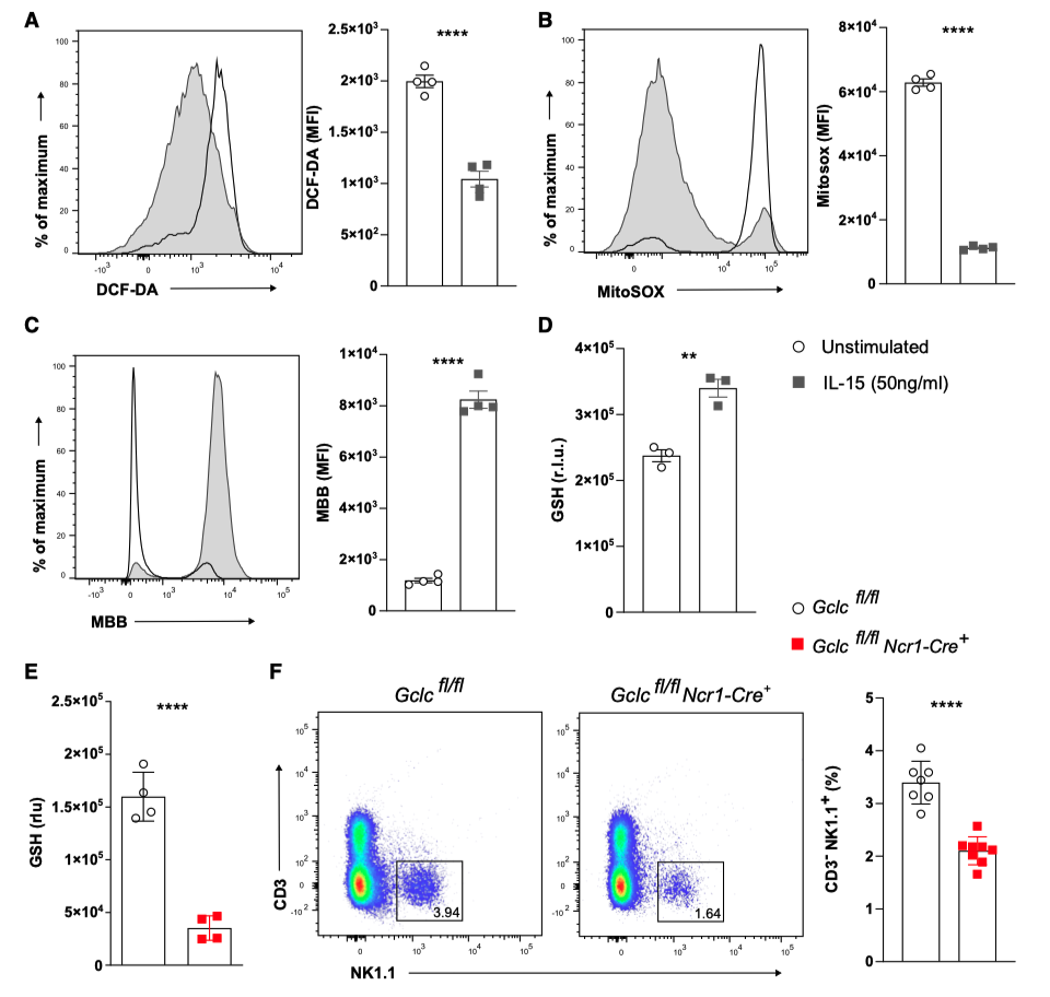
To investigate the relationship between redox equilibrium and IL-15-driven activation of NK cells, the authors stimulated the spleen NK cells of C57BL/6J wild-type (WT) mice for 24 hours, and measured the changes in intracellular ROS levels using the fluorescent probe DCF-DA.
Compared with unstimulated NK cells, the ROS level on the cell membrane of NK cells stimulated by IL-15 decreased significantly (Figure 1A), and the accumulation of ROS in the mitochondria also decreased (measured using MitoSOX) (Figure 1B).
ROS is crucial for cell activation and can regulate the cytotoxicity of NK cells.
The NK cells induced by IL-15 would increase the production of intracellular sulfhydryl compounds 27, which was manifested by a higher fluorescence of monobromomethylbenzene (MBB) (Figure 1C).
Under the stimulation of IL-15, the level of glutathione in NK cells also increases (Figure 1D).
Suppose that glutathione can regulate the activation of NK cells mediated by IL-15.
To investigate the role of glutathione in the homeostasis and function of NK cells, the authors used Gclcfl/fl mice, in which the flanking region of the catalytic subunit site of glutamine-cysteine ligase (Gclc) contains loxP elements.
These mice were crossed with Ncr1-Cre+ mice, in which the Cre recombinase gene was expressed under the control of the Ncr1 promoter, and the Ncr1 promoter is largely restricted to NK cells.
The frequency of NK cells in the spleen of Gclcfl/fl Ncr1-Cre+ mice was significantly reduced, indicating that GSH is involved in the balance of NK cells (Figure 1F).
The final characteristics of NK cell maturation and differentiation are the sequential loss of CD27.
The CD11b expression changes from immature (CD11b- CD27+) to intermediate (CD11b+ CD27+) and finally to mature (CD11b+ CD27-) in the NK cell subpopulation.
Consistent with the decreased frequency of NK cells in the spleen, the abundance of NK cells in the spleen of Gclcfl/fl Ncr1-Cre+ mice, especially CD11b+ CD27-NK cells, also decreased.
The T cells of naiveGclc-deficient mice remained unaffected.
These results indicate that glutathione can regulate the redox balance of IL-15-activated NK cells and maintain the balance of NK cells in the periphery.
Gclc ablation will damage metabolism of NK cells
Although the intracellular level of thiols was relatively low, compared with the control group of NK cells, the ROS levels on the cell membrane and mitochondria of Gclc-deficient spleen NK cells did not change during homeostasis.
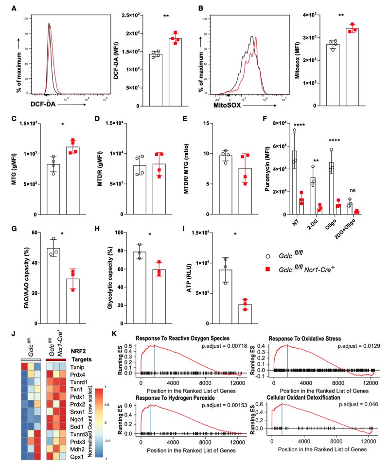
Compared with the NK cells of the control group mice, the spleen NK cells of Gclcfl/fl Ncr1-Cre+ mice showed a significant increase in the accumulation of cell membrane ROS and mitochondrial superoxide when stimulated by IL-15 (Figures 2A and 2B).
Excessive ROS is detrimental to the metabolism and function of activated NK cells.
Compared with the control NK cells, the IL-15-activated Gclc-deficient NK cells showed an increase in mitochondrial mass, but the membrane potential remained unchanged (Figures 2C and 2D).
Compared with the control NK cells, the NK cells lacking Gclc showed a trend of decreased ratio of mitochondrial activity to mitochondrial mass (Figure 2E).
This means that the absence of Gclc interferes with the mitochondrial capacity of NK cells.
The NK cells activated by IL-15 rely on oxidative phosphorylation and glycolysis to exert their effector functions. Given the impaired mitochondrial function, the authors hypothesized that the deficiency of glutathione would inhibit the metabolism of NK cells.
To assess the metabolic status of Gclc-deficient NK cells, the authors used flow cytometry to measure protein synthesis and incorporated puromycin as an alternative for energy consumption.
This method required a significantly smaller number of cells than the commonly used cells for extracellular flux analysis in the hippocampus, enabling the authors to examine the metabolic status of the scarce Gclc-deficient NK cells.
The IL-15-activated control group and Gclcfl/fl Ncr1-Cre+NK cells were either untreated or treated with oligomycin and/or 2-deoxyglucose (2-DG). Oligomycin and/or 2-DG respectively inhibited oxidative phosphorylation and glycolysis.
Compared with the control group, the NK cells with Gclc deficiency showed significantly weaker glycolytic and OXPHOS responses to IL-15 (Figure 2F).
Both oxidative phosphorylation and glycolysis need to be blocked simultaneously in order to reduce the energy capacity of the control group NK cells to the level observed in the Gclc-deficient NK cells (Figure 2F).
The deficiency of GSH in NK cells also reduces the ability of fatty acid and amino acid oxidation as well as glycolysis (Figures 2G and 2H).
The ATP levels in activated NK cells lacking Gclc were also significantly lower than those in control NK cells (Figure 2I).
These results indicate that glutathione is crucial for the metabolic activation of NK cells.
To analyze the contribution of glutathione to the transcriptional response during IL-15-driven NK cell activation, the authors performed RNA sequencing on control and Gclc-deficient NK cells to maintain the redox balance.
This is consistent with the author's observations, which show that IL-15 increases the synthesis of glutathione and sulfides, while reducing ROS in cell membranes and mitochondria (Figures 1A-1D).
IL-15-activated Gclc-deficient NK cells significantly increased the expression of antioxidant genes, many of which are controlled by NRF2, and NRF2 is the main regulator of the antioxidant response (Figure 2J).
This is reflected in the Gene Ontology (GO) terms, such as the expressions of terms like "response to ROS", "response to oxidative stress", "response to hydrogen peroxide", and "cell antioxidant detoxification" being significantly enriched (Figure 2K).
These results indicate that after the reduction of Gclc, the metabolic activation induced by IL-15 leads to an increase in ROS production and excessive accumulation.
This, in turn, enhances the NRF2-dependent antioxidant response transcription that is independent of the glutathione synthesis mechanism.
In NK cells lacking Gclc, the activation of alternative antioxidant genes cannot replace glutathione (Figures 2F-2I).
Glutathione plays an indispensable and non-redundant role in the metabolic pathway of NK cells.
Glutathione mediates IL-15-driven mTOR activation in NK cells
IL-15 can stimulate NK cells to express nutrient transporters, in order to meet the increased energy demands.
The author considered whether the deficiency of Gclc would impair the metabolic regulatory factors necessary for the activation of NK cells.
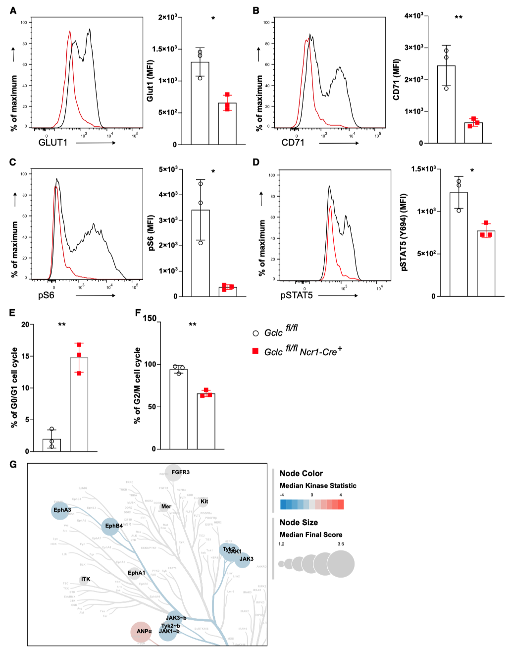
The NK cells lacking Gclc showed a significant reduction in the expression of glucose transporter GLUT1 (Figure 3A) and transferrin receptor CD71 (Figure 3B).
The expression of these transporters is regulated by the mTOR signaling pathway.
The mTOR signaling controls cell proliferation and the acquisition of cytotoxicity, and consistent with the reduced metabolic activity is that NK cells lacking Gclc exhibit a decrease in the phosphorylation of one of the downstream targets of mTOR, S6 (Figure 3C).
Whether the functions of NK cells require the continuous presence of GSH still needs to be studied.
NK cells were treated with the pharmacological antagonist of γ-glutamylcysteine synthetase (GCL) - L-dithionitroimine (BSO) to rapidly remove glutathione.
Consistent with the data obtained by the author from Gclc ablation, acute GCL inhibition significantly reduced the level of sulfhydryl in IL-15-activated WT NK cells.
The levels of phosphorylated mTOR (pmTOR) and pS6 also decreased. This indicates that the activation of mTOR in NK cells requires glutathione.
IL-15 and STAT5 phosphorylation simultaneously trigger mTOR activation, and STAT5 phosphorylation plays a crucial regulatory role in the survival, development and effector function of NK cells.
The STAT5 signal is associated with the progression of the cell cycle through the upregulation of the D-type cell cycle protein.
The NK cells lacking Gclc remained in the G0/G1 phase of the cell cycle (Figure 3E), which led to a decrease in the proportion of cells in the G2/M phase (Figure 3F).
The coordinated progression of STAT5 signal transduction and cell cycle induced by IL-15 requires Glutathione.
The weakened mTOR and STAT5 signal transduction indicates that the deficiency of Gclc extensively impairs the growth of NK cells.
The kinase activity in NK cells of the control group stimulated by IL-15 and the Gclc-deficient group was investigated using Pamgene kinase profiling analysis.
NK cells lacking glutathione exhibited a specific reduction in JAK activity (Figure 3G).
This indicates that glutathione can specifically promote the activation of JAK upstream of STAT5.
The author tested whether glutathione was necessary during the early stages of IL-15 stimulation (30, 60 and 120 minutes).
The levels of pmTOR, p-STAT5 and pS6 in NK cells lacking Gclc were comparable to those in control NK cells, except that pS6 slightly decreased at 30 minutes.
Glutathione is largely indispensable for the initial IL-15 signal transduction.
Glutathione is crucial for the IL-15-induced STAT5 and mTOR signal transduction, and STAT5 and mTOR signal transduction are necessary for the metabolism and proliferation of NK cells.
deficiency of glutathione will restrict growth and cytotoxicity of NK cells
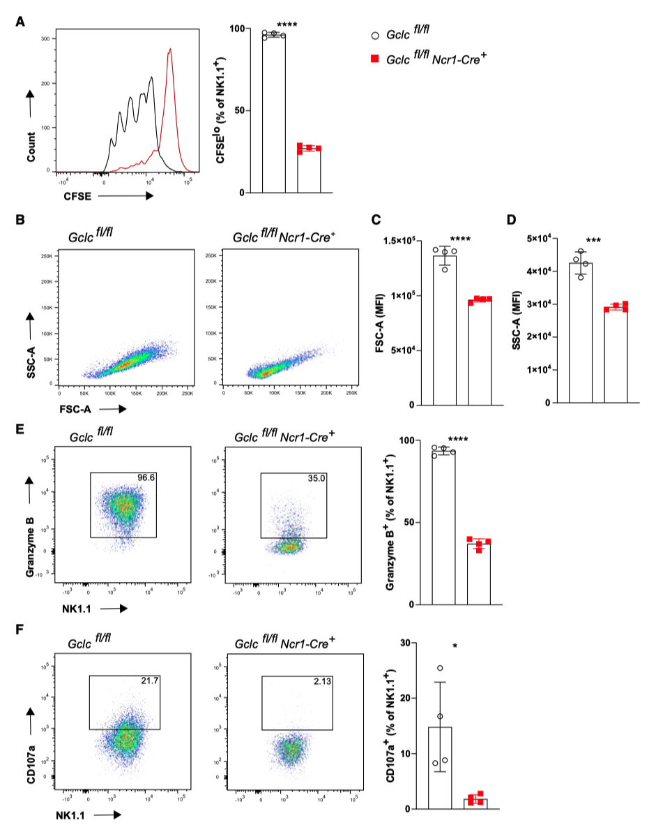
The proliferation and growth of NK cells require Gclc. Consistent with the reduction of STAT5 and mTOR signals, NK cells lacking Gclc cannot proliferate in response to IL-15 stimulation (Figure 4A).
Compared with the control cells, the NK cells lacking Gclc showed a significant reduction in cell volume and a decrease in granularity after activation (Figures 4B-4D).
NK cells with Gclc deficiency upregulated genes associated with reduced proliferation.
The genes of inhibitory and activating NK cell receptors have also been downregulated.
These results indicate that glutathione can regulate the growth and activation of NK cells.
The NK cells lacking Gclc produced less granzyme B, CD107a (Figures 4E and 4F), and IFN-γ when activated by IL-15. This indicates that glutathione is essential for the maturation of NK cell function.
Does glutathione also have the ability to regulate the activation of NK cells induced by other cytokines (such as IL-2, IL-21, or IL-12/18)?
Compared with the control NK cells, the NK cells lacking Gclc showed a significant reduction in the expression of granzyme B upon stimulation with IL-15 and IL-12/18.
These results indicate that glutathione is crucial for the maturation of NK cell function induced by IL-15.
Glutathione in NK cells is crucial for inhibiting T cell responses during acute viral infections
The role of NK cells in antiviral immunity includes their cytotoxicity towards virus-exposed cells and their ability to regulate the immune response.
In viral infections, such as lymphocytic choriomeningitis virus (LMCV) and human hepatitis B virus, NK cells play a crucial regulatory role in the activity of dendritic cells and T cells during the infection process.
Depleting NK cells using monoclonal antibodies can enhance antigen presentation and increase the production of IFN-γ by CD4+ and CD8+ T cells.
To evaluate the function of NK cells with Gclc deficiency in the body's antiviral immunity, the authors treated the control group and the Gclcfl/fl Ncr1-Cre+ group of mice with a high dose (2 × 10^6 plaque-forming units [PFUs]) of the LCMV-WE strain.
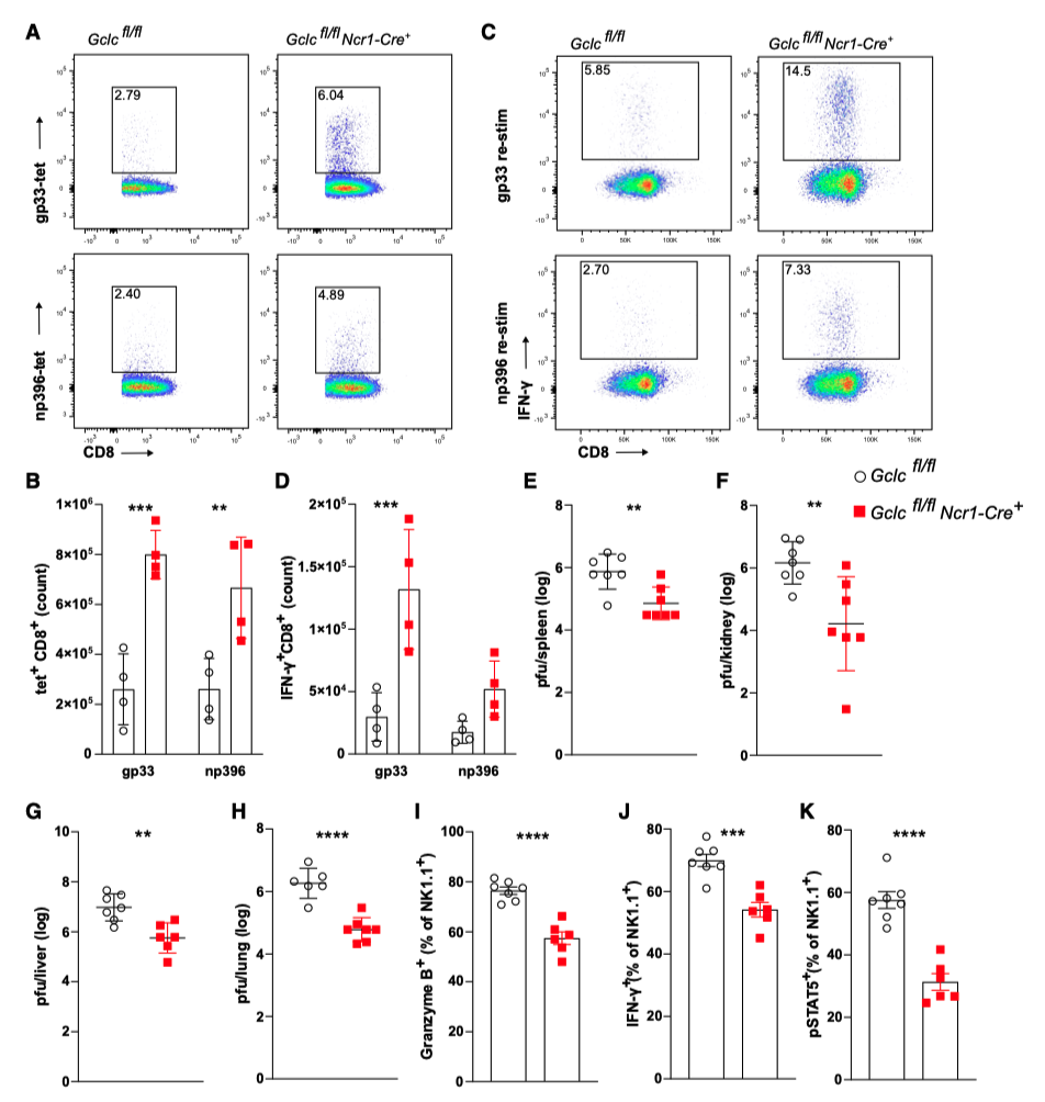
Eight days after infection, the author evaluated the quantity and function of LCMV-specific T cells, and used the major histocompatibility complex (MHC) tetramers presenting the viral glycoprotein (gp) 33 and nuclear protein (np) 396 to detect these cells.
Consistent with the weakened function of NK cells with Gclc defects, the frequency and quantity of gp33+ and np396+ CD8+ T cells in the spleen and liver of Gclcfl/fl Ncr1-Cre+ mice infected with LCMV were increased compared to those of the control group in the same litter (Figures 5A, 5B).
This is related to the increased production of IFN-γ by virus-specific T cells in the spleen and liver (Figures 5C, 5D).
Compared with the control group, the viral titers in the spleen, liver, lungs and kidneys of Gclcfl/fl Ncr1-Cre+ mice were significantly reduced (Figures 5E-5H).
Compared with the control group, the levels of liver enzymes such as aspartate aminotransferase (AST), alanine aminotransferase (ALT), and lactate dehydrogenase (LDH) in the serum of Gclcfl/fl Ncr1-Cre+ mice were lower, indicating that liver damage was alleviated.
In viral infections, NK cells can exert cytotoxic effects on T cells, thereby suppressing the antiviral response of T cells.
In fact, compared with the control group mice, the cytotoxicity of spleen cells from Gclcfl/fl Ncr1-Cre+ mice infected with LCMV was significantly reduced against YAC-1 cells.
Consistent with this, on the second day after LCMV-WE infection, NK cells with Gclc deficiency showed decreased expression of granzyme B and IFN-γ (Figures 5I and 5J).
This is related to the increased expression of LAG3 and PD1 (Figure 5G) and the decreased phosphorylation of STAT5 (Figure 5K).
These results indicate that glutathione supports the function of NK cells in inhibiting the antiviral T cell response.
Glutathione supports control of metastatic tumor spread mediated by NK cells in vivo
NK cells play a crucial role in the immune response against tumors.
The tumor microenvironment interferes with the metabolism of NK cells, weakening their anti-tumor function. ROS also limits the infiltration of NK cells into tumors.
However, how glutathione promotes the anti-tumor function of NK cells remains to be explored.
The control group and Gclcfl/fl Ncr1-Cre+ mice were subcutaneously injected with B16-F10 melanoma cells.
There was no difference in the growth of primary tumors and tumor weight between the two groups of mice.
The author also treated the Gclcfl/fl and Gclcfl/fl Ncr1-Cre+ mice with the more immunogenic MC-38 colon cancer cell line.
Similar to the B16-F10 tumors, the growth of MC-38 tumors in both groups of mice was comparable.
These results indicate that in these primary tumor models, glutathione in NK cells is indispensable for anti-tumor immunity.
The author used the B16-F10 melanoma lung metastasis model to assess the impact of glutathione deficiency on the anti-metastasis function of NK cells.
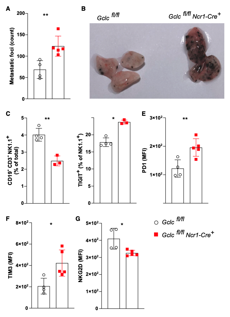
The author observed that, compared with the control mice, the lung tumor burden of Gclcfl/fl Ncr1-Cre+ mice was significantly increased (Figures 6A and 6B).
This is related to the lower infiltration of NK cells in the lungs (Figure 6C).
Compared with the control group of NK cells, the tumor-infiltrated Gclc deficient NK cells increased the expression of inhibitory receptors TIGIT, PD-1 and TIM-3, and decreased the expression of activating receptor NKG2D (Figures 6D-6G).
It is worth noting that, compared with the control group mice, the frequency of T cells or the percentage of CD8+ T cells in the tumor-infiltrating lymphocyte component of Gclcfl/fl Ncr1-Cre+ mice did not change.
The author used Rag1-/- Gclcfl/fl Ncr1-Cre+ mice to investigate the anti-metastasis function of Gclc-deficient NK cells.
These mice could be used to assess the anti-tumor function of Gclc-deficient NK cells without the involvement of adaptive immune cells (such as T cells).
After intravenous injection of B16-F10 cells, the number of metastatic lung nodules observed in Rag1-/- Gclcfl/fl Ncr1-Cre+ mice was significantly higher than that in the Rag1-/- Gclcfl/fl control group.
This indicates that glutathione can regulate the anti-metastasis function of NK cells, and this is independent of the involvement of T cells.
These results indicate that glutathione is crucial for the inhibition of tumor metastasis mediated by NK cells.


